Xiao-Bin Peng,
Gao-Jue Wu,
Xiao-Yu Wang,
Xue-Jun Tang,
Lei Gong ![]()
For correspondence:- Lei Gong Email: gonglei186@hotmail.com Tel:+8651066681222
Received: 15 September 2015 Accepted: 7 September 2016 Published: 31 October 2016
Citation: Peng X, Wu G, Wang X, Tang X, Gong L. Bryostatin I inhibits growth and proliferation of pancreatic cancer cells via suppression of NF-κB activation. Trop J Pharm Res 2016; 15(10):2071-2076 doi: 10.4314/tjpr.v15i10.3
© 2016 The authors.
This is an Open Access article that uses a funding model which does not charge readers or their institutions for access and distributed under the terms of the Creative Commons Attribution License (http://creativecommons.org/licenses/by/4.0) and the Budapest Open Access Initiative (http://www.budapestopenaccessinitiative.org/read), which permit unrestricted use, distribution, and reproduction in any medium, provided the original work is properly credited..
Purpose: To evaluate the effect of bryostatin I on proliferation of pancreatic cancer cells as well as tumor growth in mice tumor xenograft model.
Methods: Activation of NF-κB was evaluated by preparing nuclear material extract using nuclear extract kit (Carlsbad, CA, USA) followed by enzyme-linked immunosorbent assay (ELISA). Mice were injected with 3 x 10⁵ MIApaCa 2 cells in 100 μL volume of PBS. The animals in the treatment group were injected with 50 µg/kg of bryostatin 1 daily for 1 month in the morning whereas those in the untreated group received an equal volume of normal saline.
Results: Treatment of the MIApaCa 2 cells with bryostatin I caused a significant reduction in the activity of NF-κB in nucleoplasm (p = 0.0002). The increase in the concentration of bryostatin I from 10 to 50 µM reduced MIApaCa 2 cell proliferation from 87 to 26 %. Bryostatin I treatment also led to increase in the proportion of cells in M1 phase with subsequent reduction in sub-G1 phase of cell cycle. Examination of the cell lysates revealed a higher ex
Conclusion: Bryostatin I inhibits growth and proliferation of pancreatic cancer through inhibition of NF-κB ex
Introduction
Pancreatic cancer is one of the most aggressive and common causes of deaths caused by cancer with a five year survival rate of less than 4 % [1,2]. The characteristic features of the pancreatic carcinoma cells include high rate of proliferation and rapid invasive potential to adjacent tissues [3]. Pancreatic cancer because of no prominent symptoms is usually detected in its advanced stage which is a major hindrance to treatment strategies [4].
It is reported that in the regulation of cellular processes including, apoptosis, inflammation, and oncogenesis NF-κB has a vital role [5,6]. Therefore, suppression of NF-κB expression by various strategies has been found to be of great therapeutic importance for the treatment of cancers [7,8]. Enhanced expression of NF-κB was shown to correlate with aggressiveness of pancreatic cancer cell metastasis [9,10]. Activation of the factors involved in apoptosis has a great impact on the inhibition of the cancer cell proliferation and in turn carcinoma treatment [11]. NF-κB is found in the cell cytoplasm in its inactive form, p50-p65 complex and p65-p105. Activation of NF-κB is induced by the phosphorylation of IκBα which then results in translocation of NF-κB to nucleus. Penetration of NF-κB into the nucleus causes expression of the genes which initiate various activities such as induction of apoptosis and inflammation [12,13].
Phytochemical investigation of the Bugula neritina and marine bryozoa has led to the isolation of 20 macrocyclic lactones known as bryostatins. Screening of these lactones against leukemia cell lines showed that these compounds possess potent cytotoxic activities [14]. Bryostatin I the most active compound is currently in phase II clinical trials for the treatment cancers [15-17]. In addition, bryostatin I treatment has been shown to promote the growth and proliferation of bone marrow progenitor cells [17]. Thus, bryostatin I treatment has overcome the disadvantages associated with the application of dangerous ionizing radiations [18]. The present study was aimed to investigate the effect of bryostatin I on pancreatic cancer cell proliferation and tumor xenograft mice model.
Methods
Reagents
Bryostatin 1 and dimethyl sulphoxide (DMSO) were obtained from Sigma-Aldrich (St. Louis, MO, USA). Bryostatin 1 was dissolved in DMSO and stored under inert atmosphere at -40 oC.
Cell line and culture
Human pancreatic carcinoma cell line, MIApaCa 2 was obtained from the American Type Culture Collection (Rockville, MD, USA). The cells were cultured in RPMI 1640 medium (Nissui Pharmaceutical Co., Ltd., Tokyo, Japan) containing 10 % heat inactivated fetal bovine serum (JRH Biosciences, Lenexa, KS, USA), and antibiotics. The cells were maintained in a humidified atmosphere of 5 % CO2 at 37 oC.
Animals
Thirty 8-week old mice, (BALBc nu/nu) were obtained from CLEA Japan Incorporated (Tokyo, Japan). All experiments involving animals were conducted in accordance with the National Institute of Health Guide for the Care and Use of Laboratory Animals [19]. All animal experiments were approved by the Ethics Committee of Wuxi No. 2 Hospital Affiliated to Nanjing Medical University (protocol no. FGS20131108). The mice were acclimatized to the laboratory atmosphere one week before the start of actual experiment. All the mice were caged and housed in the rooms with 12 h light and dark cycle at 25 oC temperature with free access to food and water. All the experiments on animals followed the National Institutes of Health criteria for the care and use of laboratory animals. The study was also approved by the Laboratory Animal Care Committee of Sun Yat-sen University (Guangzhou, China).
Quantitative analysis of NF-κB activity
MIApaCa 2 cells were treated with 10 - 50 µM of bryostatin 1 for 12, 24 and 48 h and then analyzed for the expression of NF-κB p65. Following incubation, nuclear extract kit (Carlsbad, CA, USA) was used to prepare the extract of the nuclear material as per the manual instructions. The nuclear extract of the cells was then analyzed by enzyme-linked immunosorbent assay (ELISA) kit (TransAM™ NF-κB; Active Motif). For this purpose, 5 μg samples of the nuclear material were incubated with oligonucleotide containing NF-κB p65-binding sequence for a period of 45 min at 30 oC in a microwell. The nucleoplasm samples were then incubated with rabbit anti-NF-κB p65 antibodies (Qiagen, TX, USA) for 45 min at 30 oC, followed by peroxidase-conjugated goat anti-rabbit IgG (Qiagen, TX, USA) for 45 min at 30 oC. Tetramethylbenzidine reaction was used for the visualization of peroxidase activity and the optimal density was measured at 465 nm.
Cell proliferation assay
The standard 3 (4, 5 dimethylthiazol 2 yl) 2,5 diphenyltetrazolium bromide assay was used for the analysis of inhibition of proliferation in pancreatic carcinoma cells. The cells were treated with bryostatin 1 at various concentrations (10, 20, 30, 40 and 50 μM) for various time periods or with DMSO alone as the control. The density of the bryostatin 1- and DMSO treated cells was compared to determine the rate of cell proliferation inhibition by bryostatin 1.
Western blot analysis
MIApaCa 2 cells after incubation with bryostatin 1 were washed with PBS and treated with lysis buffer (Beyotime). The concentration of the proteins in the cell lysates was measured by Bradford method using bovine serum albumin as standard. 5 mg samples of the proteins were loaded into 10 % SDS-polyacrylamide gels and subjected to electrophoresis using constant voltage. The proteins were then transferred onto PVDF membranes (Millipore, Bedford, MA, USA). The non-specific sites in the membrane were blocked by incubation with blocking buffer (5 % skim milk in TBS-T) for 3 h at room temperature. The membrane was then incubated with pro- and cleaved caspase-8, IκB· and phosphorylated IκB· monoclonal antibodies (Santa Cruz Biotechnology, Inc.) at 4 oC overnight. The membranes were washed three times with TBS-T and then incubated with a secondary antibody (Santa Cruz Biotechnology, Inc.) at room temperature for 1 h. The immunoreactive bands were visualized using enhanced chemiluminescence kits (Pierce Biotechnology Inc., Rockford, IL, USA). Blots were stained with anti-β-actin or -GAPDH antibody (Santa Cruz Biotechnology, Inc.) as an internal control for the amounts of target proteins.
Cell cycle analysis
MIApaCa 2 cells were distributed at a density of 3 x 10⁶ cells per well in 6-well plates at 37 oC and incubated with bryostatin 1. After incubation for 48 h, the cells were washed with PBS, resuspended with 200 μL RNase A (1 mg/mL) and PI (200 μg/mL) (Beyotime, Haimen, China) for staining the DNA content. Following incubation for 30 min at room temperature in the dark, the cellular DNA was analyzed by FACSCalibur flow cytometer. ModFitLT V2.0 software (both from Becton-Dickinson) was used for the analysis of the data.
Preparation of pancreatic xenograft cancer model
3 x 10⁵ MIApaCa 2 cells suspended in 100 μl volume of PBS were injected into the mice on right dorsal side of the body. The animals were then randomly divided into the two groups of 15 animals each (treatment and untreated groups). The animals in the treatment group were injected 50 µg/kg body weight doses of bryostatin 1 dialy for 1 month in the morning whereas those in the untreated group received equal volume of normal saline. Five animals from each of the group were sacrificed after 30, 60 and 90 days following administration of MIApaCa 2 cells. The tumors mass from each of the animal was extracted, washed and weighed.
Statistical analysis
The results are expressed as mean ± standard deviation (SD). For statistical analysis, SPSS 16.0 software was used. Comparison of the results was performed using one-way analysis of variance (ANOVA) and Student-Newman-Keuls test. Differences were considered statistically significant at p < 0.05.
Results
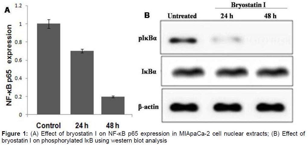
Effect of bryostatin I on NF-κB activity
Treatment of the MIApaCa 2 cells with bryostatin I caused a significant reduction in the activity of NF-κB in nucleoplasm compared to untreated cells (p = 0.0002, A). Bryostatin I treatment markedly inhibited the activation of IκB in MIApaCa 2 cells after 48 h compared to untreated cells (B). It was observed that the level of activated IκB in bryostatin I treated MIApaCa 2 cells was significantly lower than those in untreated cells.
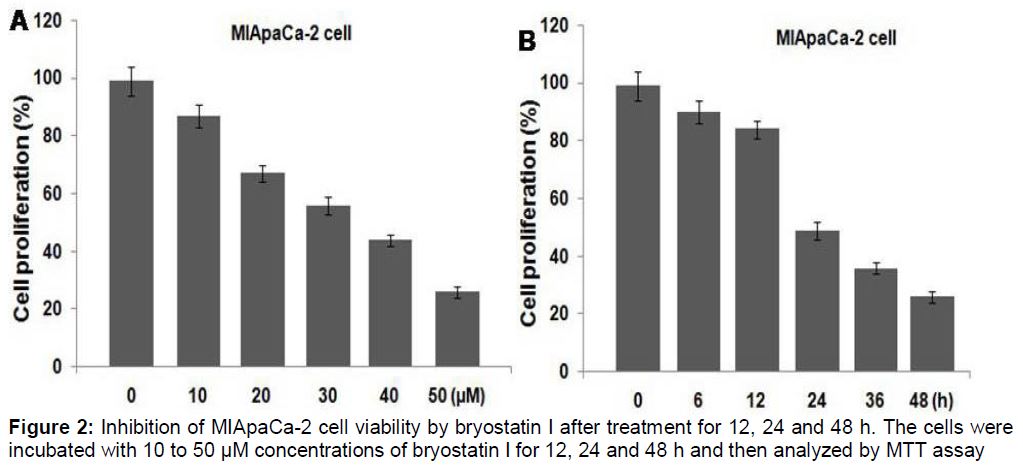
Bryostatin I Inhibits proliferation of MIApaCa‑2 cells
The results from MTT assay revealed that bryostatin I exhibited concentration and time dependent inhibitory effects on the proliferation of MIApaCa‑2 cells. Increase in concentration of bryostatin I from 10 to 50 µM reduced the percentage of MIApaCa‑2 cell proliferation from 87 to 26 % (). The rate of proliferation in MIApaCa‑2 cells on treatment with 50 µM bryostatin I after 12, 24 and 48 h was found to be 91.23 ± 6.11, 43.89 ± 3.45 and 26.14 ± 2.67 %, respectively ().
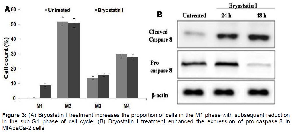
Bryostatin I induced apoptosis in MIApaCa‑2 cells
Bryostatin I treatment led to the increase in proportion of cells in M1 phase compared to the untreated cells (A). However, the proportion of cells in sub-G1 phase of cell cycle was reduced by bryostatin I treatment. Examination of the cell lysates revealed higher expression level of cleaved caspase-8 in the bryostatin I treated MIApaCa‑2 cells compared to untreated cells (B).
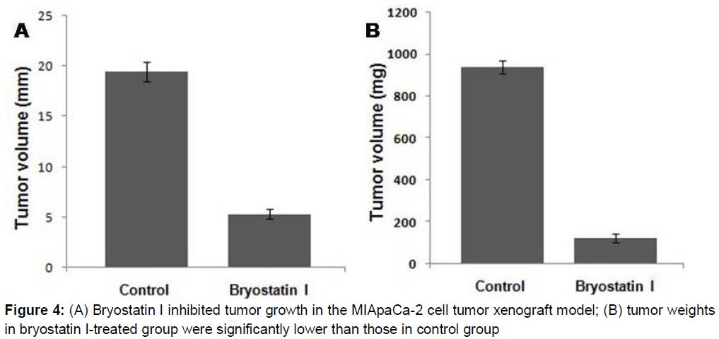
Bryostatin I treatment inhibits tumor growth in vivo
Comparison of the tumor growth in bryostatin I treated and untreated mice after 2 months revealed a significantly lower tumor volume in treatment group than in untreated group (a). The average tumor volumes in the treatment and untreated groups were found to be 5.34 ± 2.16 and 19.45 ± 5.71 mm, respectively after 2 months of the treatment period (p < 0.0002) (A). The average weight of the tumors in the treatment and untreated groups were 123.67 ± 22.56 and 939.14 ± 213.51 mg, respectively after 2 months of the treatment (B).
Discussion
The present study investigates the effect of bryostatin I on proliferation of pancreatic cancer cells as well as tumor growth in mice tumor xenograft model. The study revealed that bryostatin I treatment results apoptosis in pancreatic carcinoma cells and inhibits growth of tumor in vivo by preventing the expression of NF-κB. The genes associated with the expression of proteins involved in the cellular processes, including apoptosis and inflammation are regulated by NF-κB expression. It is known that activation of NF-κB is inhibited during the process of inflammation [20,21]. The chemotherapeutic inhibition of NF-κB expression prevents proliferation, invasion and metastasis of cells in various types of cancers [22-24]. Although, a number of agents have been identified which play an important role in the inhibition of NF-κB expression. However, most of these agents induce harmful side effects [25]. Therefore, the discovery of the molecules which can inhibit the expression of NF-κB without side effect is highly desired. Our results from the current study revealed that bryostatin I treatment inhibited the expression of NF-κB in the pancreatic carcinoma cells. The level of activated IκB in MIApaCa 2 cells was reduced markedly on treatment with bryostatin I. Inhibition of carcinoma cell proliferation by the use of various agents plays important value in the treatment of cancer. The present study revealed that bryostatin I treatment caused a significant inhibition in the rate of proliferation in pancreatic carcinoma cells. Bryostatin I treatment in pancreatic cells resulted cell cycle arrest by increasing proportion of cells in M1 phase and subsequent reduction in sub-G1 phase of cell cycle. Western blot analysis showed significantly higher expression level of cleaved caspase-8 in the bryostatin I treated Panc-1 cells compared to untreated cells. Thus, bryostatin I induced apoptosis in pancreatic carcinoma cells through caspase-dependent pathway. Comparison of the tumor growth in bryostatin I treated and untreated mice after 2 months revealed a significantly lower tumor volume in treatment group than in untreated group.
Conclusion
The findings of this study indicate that bryostatin I induces apoptosis in pancreatic carcinoma cells and inhibits tumor growth in vivo through down-regulation of NF-κB expression. Therefore, bryostatin I may be of therapeutic value for the treatment of pancreatic cancer.
Declarations
Acknowledgement
References
Archives


News Updates